pelvis model human skeleton model specimen hip skeleton anatomy medical tool school used 1 1 pubis skeletal educational

This Book is a... 3D Human Body!
Explore your insides in 3D detail! Press out the cleverly shaped, chunky pages to reveal a 3D model of the human body. Discover astonishing facts about every layer, from skin to skeleton, and see how they all fit together. This combined fact book and play model is the perfect interactive introduction to the world inside every body.
1442 Руб.
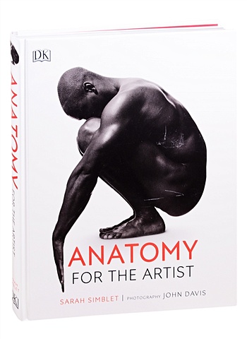
Anatomy for the Artist
Unlock your inner artist and learn how to draw the human body in this beautifully illustrated art book by celebrated artist and teacher Sarah Simblet. This visually striking guide takes a fresh approach to drawing the human body. A combination of innovative photography and drawings, practical life-drawing lessons, and in-depth explorations of the body’s surface and underlying structure are used to reveal and celebrate the human form. Combining specially-commissioned photographs of models with historical and contemporary works of art and her own dynamic life drawing, Sarah leads us inside the human body to map its skeleton, muscle groups, and body systems. Detailed line drawings superimposed over photographs reveal the links between the body’s appearance and its construction. Six drawing classes show how to observe different parts of the body and give expert guidance on how to draw them. Inspirational master classes on famous works, ranging from a Michelangelo study to a Degas painting, show how artists have depicted the human body over the centuries. Each master class includes a photograph of a model holding the same pose as in the painting, to highlight details of anatomy and show how the artist has interpreted them. Understanding anatomy is the key to drawing the human body successfully. As well as being the perfect reference, Anatomy for the Artist will inspire you to find a model, reach for your pencil, and start drawing.
1800 Руб.

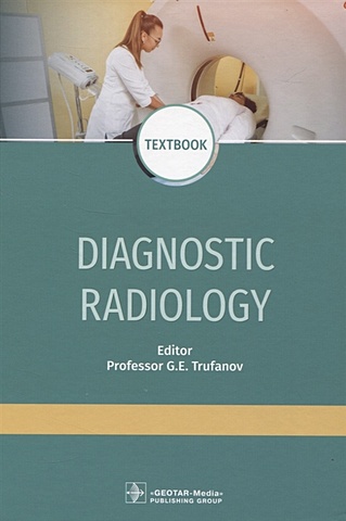
8Pcs Skeleton Model Wholesale Learn Aid Anatomy Art Sketch Halloween Flexible Human Anatomical Anatomy Bone
296.6 Руб.
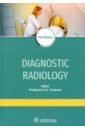
Труфанов Геннадий Евгеньевич, Акиев Рустам Магомедович, Алексеев Кирилл Николаевич Diagnostic radiology. Textbook
The textbook contains the basic principles of radiation diagnosis of injuries and diseases of human organs and systems, the characteristics of all methods of radiation diagnosis with a description of the physical principles of imaging. The radiation anatomy of human organs and systems, as well as the features of research, are described from the modern point of view. The possibilities of radiation research methods in the diagnosis of diseases and injuries of various organs and systems are reviewed. The radiological semiotics of injuries and the most common diseases of the skeletal system, organs of the thoracic cavity, abdomen, pelvis, as well as the brain and spinal cord are described in detail. At the end of each section, the indications for the use of a particular method for examination of various organs and systems are represented accurately. The textbook was composed in compliance with the modern requirements of the Federal State Educational Standards of Higher Education and is intended for the medical students of the departments “Radiation Diagnosis”, “Radiation Diagnosis and Radiation Therapy”, “Radiation Diagnosis (Radiology)”; may be useful in the implementation of the principal educational program of the postgraduate professional education in the training of highly qualifi ed specialists in the discipline 08.31.09 “Radiology”. 3-е издание, переработанное и дополненное
3003 Руб.
Труфанов Г. (ред.) Diagnostic radiology
The textbook contains the basic principles of radiation diagnosis of injuries and diseases of human organs and systems, the characteristics of all methods of radiation diagnosis with a description of the physical principles of imaging. The radiation anatomy of human organs and systems, as well as the features of research, are described from the modern point of view. The possibilities of radiation research methods in the diagnosis of diseases and injuries of various organs and systems are reviewed. The radiological semiotics of injuries and the most common diseases of the skeletal system, organs of the thoracic cavity, abdomen, pelvis, as well as the brain and spinal cord are described in detail. At the end of each section, the indications for the use of a particular method for examination of various organs and systems are represented accurately. The textbook was composed in compliance with the modern requirements of the Federal State Educational Standards of Higher Education and is intended for the medical students of the departments "Radiation Diagnosis", "Radiation Diagnosis and Radiation Therapy", "Radiation Diagnosis (Radiology)"; may be useful in the implementation of the principal educational program of the postgraduate professional education in the training of highly qualifi ed specialists in the discipline 08.31.09 "Radiology".
2805 Руб.
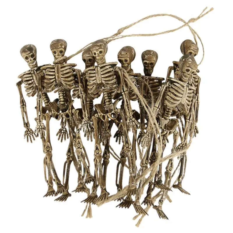
8 Pcs Human Skeleton Model Human Skeleton Set With Hangings Rope Props Trick Supplies Life Size Skeleton With Hangings Rope For
4942.95 Руб.
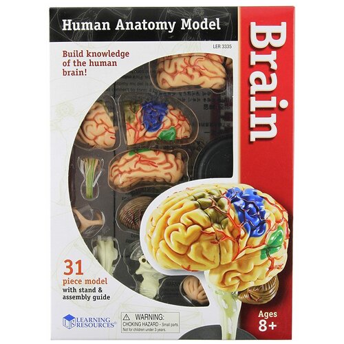
Набор Learning Resources Human Anatomy Model Brain, 1 эксперимент
Конструктор Learning Resources Анатомия человека Мозг (31 элемент) Непосредственная работа с элементами набора позволяет юным исследователям наглядно познакомиться с внутренним устройством человеческого мозга и понять, как работает этот сложный механизм. Набор состоит из подставки, реалистичной детализированной модели мозга, описания ключевых фактов и пошаговой инструкции по сборке с фотоиллюстрациями. В наборе 31 деталь: Мозжечок, лобная, теменная, височная доля, затылочной, мозолистое тела, ствол мозга, гиппокамп, желудочки, полосатое тело, внутренняя капсула и чечевицевидное ядро. Размер в собранном виде: 10см (без учета подставки).
6690 Руб.
Дыдыкин Сергей Сергеевич, Сеченов Иван Михайлович, Блинова Екатерина Валериевна Topographic Anatomy and Operative Surgery. Workbook. In 2 parts. Part I
This workbook on the subject «Topographic anatomy and operative surgery» was prepared by the staff of the department of operative surgery and topographic anatomy of the Sechenov University following the requirements of the federal state educational standard of higher professional education in specialties «General Medicine» and «Pediatrics». In the workbook topics are grouped by the discipline sections. Its first part contains areas devoted to the subject of topographic anatomy and operative surgery of the upper and lower extremities, head and neck. The second part includes the abdomen, pelvis and chest. This workbook is intended for students of general medicine and pediatrics departments of medical universities. The factual material underlying the workbook is contained in the textbooks «Operative Surgery and Topographic Anatomy» edited by V.V. Kovanov (1995, 2001), «Topographic anatomy and operative surgery» by A.V. Nikolaev (2007, 2009, 2015).
4006 Руб.
Дыдыкин Сергей Сергеевич, Сеченов Иван Михайлович, Блинова Екатерина Валериевна Topographic Anatomy and Operative Surgery. Workbook. In 2 parts. Part II
This workbook on the subject «Topographic anatomy and operative surgery» was prepared by the staff of the department of operative surgery and topographic anatomy of the Sechenov University following the requirements of the federal state educational standard of higher professional education in specialties «General Medicine» and «Pediatrics». In the workbook topics are grouped by the discipline sections. Its first part contains areas devoted to the subject of topographic anatomy and operative surgery of the upper and lower extremities, head and neck. The second part includes the abdomen, pelvis and chest. This workbook is intended for students of general medicine and pediatrics departments of medical universities. The factual material underlying the workbook is contained in the textbooks «Operative Surgery and Topographic Anatomy» edited by V.V. Kovanov (1995, 2001), «Topographic anatomy and operative surgery» by A.V. Nikolaev (2007, 2009, 2015).
4006 Руб.
Roberts Alice The Complete Human Body. The Definitive Visual Guide
Intricate details of all aspects of the human body down to the smallest detail - from our cells and DNA, to the largest bone in our bodies, the femur. 3D generated illustrations and medical imaging provide a close look at the body's forms and functions in physiology and anatomy, showing how the body works and its amazing systems and abilities. To understand our modern human bodies, this book first looks at our ancestors and how the evolution of Homo Sapiens shaped our anatomy. This gave us the ability to walk tall, create language, and make tools with our incredibly adapted apposable thumbs. Learn how we can see evolution in our DNA, and the functions of DNA. Read about the things you can only see with microscopes and other special imaging machines, like cell structure, motor pathways in the brain, and the inner iris. All these many parts work together to make the human body. The physiology of our body is written in clarifying detail. Learn about the organs and systems that operate within, such as the cardiovascular, digestive, and neural systems. See our elegant anatomy and read how the skeleton, muscles, and ligaments operate to allow movement. This second addition has included more detail on the joints in the hands and feet. The Complete Human Body takes you from infancy to old age showing how our body grows and changes, and what can go wrong. 2nd Edition: Enhanced and Updated This visual guide uses remarkable illustrations and diagrams to let you peek inside our complex and astounding bodies. It has been written in an easy-to-follow format, with straightforward explanations to give you the best overview of the many things that make us human. Suitable for young students who want an extra resource for school, people working in medical fields, or for anyone with a keen interest in human biology. Inside the body of the book: - The Integrated Body - Anatomy - How the Body Works - Life Cycles - Diseases and Disorders
6495 Руб.
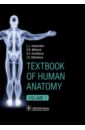
Колесников Лев Львович, Никитюк Дмитрий Борисович, Клочкова С. В. Textbook of Human Anatomy. In 3 vol. Volume 1. Locomotor apparаtus
This book provides essential facts of human anatomy for medical students. It demonstrates the basic knowledge for exam preparation and practice review of visual experiences. Plenty of clear illustrations (more than 900 pictures, radiographic and cross-sectional images) help students memorize the topics of anatomy. Modern imaging technologies allow the depiction of organs and systems in a variety of ways to gain thorough knowledge and to link to the clinical setting. The content of the book corresponds to the Federal Program for medical education. Material is divided according to systemic anatomy into three volumes. Volume 1 contains Introduction, Human development, and Locomotor apparatus.
4999 Руб.
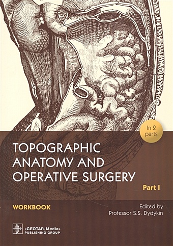
Дыдыкин С. (ред.) Topographic Anatomy and Operative Surgery. Workbook. In 2 parts. Part I
This workbook on the subject "Topographic anatomy and operative surgery" was prepared by the staff of the department of operative surgery and topographic anatomy of the Sechenov University following the requirements of the federal state educational standard of higher professional education in specialties "General Medicine" and "Pediatrics". In the workbook topics are grouped by the discipline sections. Its first part contains areas devoted to the subject of topographic anatomy and operative surgery of the upper and lower extremities, head and neck. The second part includes the abdomen, pelvis and chest. This workbook is intended for students of general medicine and pediatrics departments of medical universities. The factual material underlying the workbook is contained in the textbooks "Operative Surgery and Topographic Anatomy" edited by V.V. Kovanov (1995, 2001), "Topographic anatomy and operative surgery" by A.V. Nikolaev (2007, 2009, 2015).
2537 Руб.
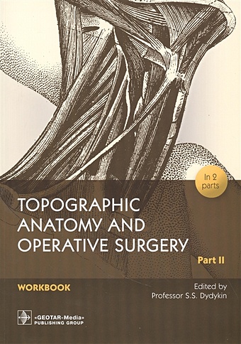
Дыдыкин С. (ред.) Topographic Anatomy and Operative Surgery. Workbook. In 2 parts. Part II
This workbook on the subject "Topographic anatomy and operative surgery" was prepared by the staff of the department of operative surgery and topographic anatomy of the Sechenov University following the requirements of the federal state educational standard of higher professional education in specialties "General Medicine" and "Pediatrics". In the workbook topics are grouped by the discipline sections. Its first part contains areas devoted to the subject of topographic anatomy and operative surgery of the upper and lower extremities, head and neck. The second part includes the abdomen, pelvis and chest. This workbook is intended for students of general medicine and pediatrics departments of medical universities. The factual material underlying the workbook is contained in the textbooks "Operative Surgery and Topographic Anatomy" edited by V.V. Kovanov (1995, 2001), "Topographic anatomy and operative surgery" by A.V. Nikolaev (2007, 2009, 2015).
2537 Руб.
Дыдыкин С. (ред.) Topographic Anatomy and Operative Surgery. Workbook. In 2 parts. Part I
This workbook on the subject "Topographic anatomy and operative surgery" was prepared by the staff of the department of operative surgery and topographic anatomy of the Sechenov University following the requirements of the federal state educational standard of higher professional education in specialties "General Medicine" and "Pediatrics". In the workbook topics are grouped by the discipline sections. Its first part contains areas devoted to the subject of topographic anatomy and operative surgery of the upper and lower extremities, head and neck. The second part includes the abdomen, pelvis and chest. This workbook is intended for students of general medicine and pediatrics departments of medical universities. The factual material underlying the workbook is contained in the textbooks "Operative Surgery and Topographic Anatomy" edited by V.V. Kovanov (1995, 2001), "Topographic anatomy and operative surgery" by A.V. Nikolaev (2007, 2009, 2015).
2537 Руб.