life size human nose anatomy model sense organ nasal cavity anatomical medical sciences educational equipment teaching
Каган Илья Иосифович, Лященко Сергей Николаевич, Мирончев Антон Олегович Topographic and clinical anatomy of the human body. The teaching aid for foreign students
Th is teaching aid is the additional source of information on topographical anatomy of regions of the human body and clinical anatomy of inner organs to the main textbook. Its English version is intended for students who take a training course in foreign faculty of the university. The teaching aid is illustrated by pictures, which are located on opening pages. On each such opening page, to the right on the odd page, there is the description of region or organ, and to the left on the even page, there is its location according to the anatomic region or organ picture. Such disposition makes the presentation of educational material more visual and makes the learning process easier.
2806 Руб.
Kagan I., Lyashchenko S., Mironchev A. Topographic and clinical anatomy of the human body: the teaching aid for foreign students
Th is teaching aid is the additional source of information on topographical anatomy of regions of the human body and clinical anatomy of inner organs to the main textbook. Its English version is intended for students who take a training course in foreign faculty of the university. The teaching aid is illustrated by pictures, which are located on opening pages. On each such opening page, to the right on the odd page, there is the description of region or organ, and to the left on the even page, there is its location according to the anatomic region or organ picture. Such disposition makes the presentation of educational material more visual and makes the learning process easier.
2240 Руб.
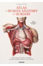
Bourgery J. M., Jacob N. M. Atlas of Human Anatomy and Surgery. The Complete
Discover one of the most comprehensive and beautifully illustrated anatomical treatises ever published. The product of more than two decades of dedication, and spanning descriptive, surgical, microscopic, and general anatomy, as well as embryology, The Atlas of Anatomy marks to this day a major achievement in medical history, and a breathtaking insight into the workings and wonder of the human body.
5049 Руб.
Human Anatomy
The definitive visual guide to human anatomy Discover fascinating facts about the human body in Human Anatomy. Did you know that your stomach is only a centimetre away from the bottom of your heart? You'll get to see all the biological wonders of the human body, often at life-size, with spectacular anatomical images showing the body's structure in incredible detail, from bone sutures to lymph nodes. Professor Alice Roberts takes you on a journey through all the body's systems, working down from the head to the toes, with a clear overview of each system. Amazing body diagrams and exhaustive annotations give you all the key details on organs and body structures. Human Anatomy is perfect for students of human biology and physiology with the quality of digital images and level of detail making it an invaluable study resource. Or if you are just curious to learn how your body works, this anatomy atlas is the perfect guide for you.
2572 Руб.
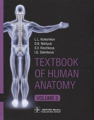
Колесников Лев Львович, Никитюк Дмитрий Борисович, Клочкова Светлана Валерьевна Textbook of Human Anatomy. Volume 3. Nervous system
This book provides essential facts of human anatomy for medical students. It demonstrates the basic knowledge for exam preparation and practice review of visual experiences. Plenty of clear illustrations (more than 900 pictures, radiographic and cross-sectional images) help students memorize the topics of anatomy. Modern imaging technologies allow the depiction of organs and systems in a variety of ways to gain thorough knowledge and to link to the clinical setting. The content of the book corresponds to the Federal Program for medical education. Material is divided according to systemic anatomy into three volumes. Volume 3 contains information about nervous system structure (Central, Peripheral, Autonomic nervous systems) and sense organs.
3630 Руб.
Le Minor Jean-Marie, Sick Henri Bourgery. Atlas of Human Anatomy and Surgery
We owe a great debt to Jean Baptiste Marc Bourgery (1797–1849) for his Atlas of Anatomy, which was not only a massive event in medical history, but also remains one of the most comprehensive and beautifully illustrated anatomical treatises ever published in any language. In 1830, having received his doctorate in medicine three years prior, Bourgery began work on his magnificent atlas in cooperation with illustrator Nicolas Henri Jacob (1782–1871), a student of the French painter Jacques Louis David. The first volumes were published the following year, but completion of the treatise required nearly two decades of dedication; Bourgery lived just long enough to finish his labor of love, but the last of the treatise’s eight volumes was not published in its entirety until five years after his death. The four parts of Bourgery’s treatise cover descriptive anatomy, surgical anatomy and techniques (exploring in detail nearly all the major operations that were performed during the first half of the 19th century), general anatomy and embryology, and microscopic anatomy. Jacob’s spectacular hand-colored, life-size lithographs are remarkable for their clarity, color, and aesthetic appeal, reflecting a combination of direct laboratory observation and illustrative research; the images are to this day unsurpassed in anatomical illustration.
3864 Руб.
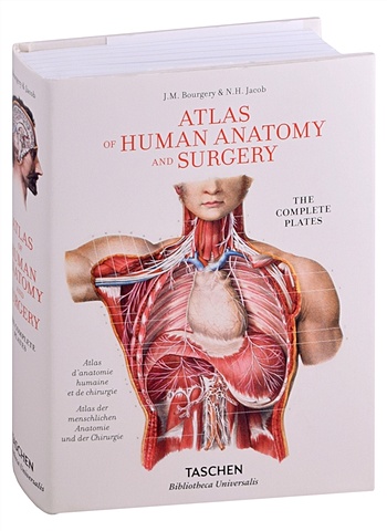
Bourgery J. M., Jacob N. H. Atlas of Human Anatomy and Surgery
We owe a great debt to Jean Baptiste Marc Bourgery (1797-1849) for his Atlas of Anatomy, which was not only a massive event in medical history, but also remains one of the most comprehensive and beautifully illustrated anatomical treatises ever published. Bourgery began work on his magnificent atlas in 1830 in cooperation with illustrator Nicolas Henri Jacob (1782-1871), a student of the French painter Jacques Louis David. The first volumes were published the following year, but completion of the treatise required nearly two decades of dedication; Bourgery lived just long enough to finish his labor of love, but the last of the treatise's eight volumes was not published in its entirety until five years after his death. The eight volumes of Bourgery's treatise cover descriptive anatomy, surgical anatomy and techniques (exploring in detail nearly all the major operations that were performed during the first half of the 19th century), general anatomy and embryology, and microscopic anatomy. Jacob's spectacular hand-colored lithographs are remarkable for their clarity, color, and aesthetic appeal, reflecting a combination of direct laboratory observation and illustrative research. Unsurpassed to this day, the images offer exceptional anatomical insight, not only for those in the medical field but also for artists, students, and anyone interested in the workings and wonder of the human body.
15147 Руб.
Bourgery J.M., Jacob N.H. Atlas of Human Anatomy and Surgery
We owe a great debt to Jean Baptiste Marc Bourgery (1797-1849) for his Atlas of Anatomy, which was not only a massive event in medical history, but also remains one of the most comprehensive and beautifully illustrated anatomical treatises ever published.Bourgery began work on his magnificent atlas in 1830 in cooperation with illustrator Nicolas Henri Jacob (1782-1871), a student of the French painter Jacques Louis David. The first volumes were published the following year, but completion of the treatise required nearly two decades of dedication; Bourgery lived just long enough to finish his labor of love, but the last of the treatise's eight volumes was not published in its entirety until five years after his death.The eight volumes of Bourgery's treatise cover descriptive anatomy, surgical anatomy and techniques (exploring in detail nearly all the major operations that were performed during the first half of the 19th century), general anatomy and embryology, and microscopic anatomy. Jacob's spectacular hand-colored lithographs are remarkable for their clarity, color, and aesthetic appeal, reflecting a combination of direct laboratory observation and illustrative research. Unsurpassed to this day, the images offer exceptional anatomical insight, not only for those in the medical field but also for artists, students, and anyone interested in the workings and wonder of the human body.
2471 Руб.

Teaching equipment, biological teaching model, juvenile human body half-body model, height 45cm
2870.49 Руб.
Колесников Лев Львович, Никитюк Дмитрий Борисович, Клочкова С. В. Textbook of Human Anatomy. In 3 vol. Volume 1. Locomotor apparаtus
This book provides essential facts of human anatomy for medical students. It demonstrates the basic knowledge for exam preparation and practice review of visual experiences. Plenty of clear illustrations (more than 900 pictures, radiographic and cross-sectional images) help students memorize the topics of anatomy. Modern imaging technologies allow the depiction of organs and systems in a variety of ways to gain thorough knowledge and to link to the clinical setting. The content of the book corresponds to the Federal Program for medical education. Material is divided according to systemic anatomy into three volumes. Volume 1 contains Introduction, Human development, and Locomotor apparatus.
4999 Руб.
Колесников Л., Никитюк Д., Клочкова С., Стельникова И. Textbook of Human Anatomy. In 3 volume. Volume 3. Nervous system. Esthesiology/Анатомия человека. Учебник на английском языке в трех томах. Том 3
This book provides essential facts of human anatomy for medical students. It demonstrates the basic knowledge for exam preparation and practice review of visual experiences. Plenty of clear illustrations (more than 900 pictures, radiographic and cross-sectional images) help students memorize the topics of anatomy.
3249 Руб.
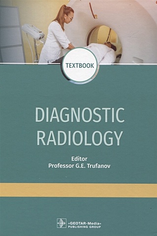
Труфанов Г. (ред.) Diagnostic radiology
The textbook contains the basic principles of radiation diagnosis of injuries and diseases of human organs and systems, the characteristics of all methods of radiation diagnosis with a description of the physical principles of imaging. The radiation anatomy of human organs and systems, as well as the features of research, are described from the modern point of view. The possibilities of radiation research methods in the diagnosis of diseases and injuries of various organs and systems are reviewed. The radiological semiotics of injuries and the most common diseases of the skeletal system, organs of the thoracic cavity, abdomen, pelvis, as well as the brain and spinal cord are described in detail. At the end of each section, the indications for the use of a particular method for examination of various organs and systems are represented accurately. The textbook was composed in compliance with the modern requirements of the Federal State Educational Standards of Higher Education and is intended for the medical students of the departments "Radiation Diagnosis", "Radiation Diagnosis and Radiation Therapy", "Radiation Diagnosis (Radiology)"; may be useful in the implementation of the principal educational program of the postgraduate professional education in the training of highly qualifi ed specialists in the discipline 08.31.09 "Radiology".
2805 Руб.