life size human kidney diseased model anatomical anatomy diseased pathological stone organ teaching supplies
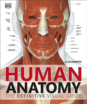
Human Anatomy
The definitive visual guide to human anatomy Discover fascinating facts about the human body in Human Anatomy. Did you know that your stomach is only a centimetre away from the bottom of your heart? You'll get to see all the biological wonders of the human body, often at life-size, with spectacular anatomical images showing the body's structure in incredible detail, from bone sutures to lymph nodes. Professor Alice Roberts takes you on a journey through all the body's systems, working down from the head to the toes, with a clear overview of each system. Amazing body diagrams and exhaustive annotations give you all the key details on organs and body structures. Human Anatomy is perfect for students of human biology and physiology with the quality of digital images and level of detail making it an invaluable study resource. Or if you are just curious to learn how your body works, this anatomy atlas is the perfect guide for you.
2572 Руб.
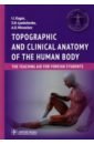
Каган Илья Иосифович, Лященко Сергей Николаевич, Мирончев Антон Олегович Topographic and clinical anatomy of the human body. The teaching aid for foreign students
Th is teaching aid is the additional source of information on topographical anatomy of regions of the human body and clinical anatomy of inner organs to the main textbook. Its English version is intended for students who take a training course in foreign faculty of the university. The teaching aid is illustrated by pictures, which are located on opening pages. On each such opening page, to the right on the odd page, there is the description of region or organ, and to the left on the even page, there is its location according to the anatomic region or organ picture. Such disposition makes the presentation of educational material more visual and makes the learning process easier.
2806 Руб.
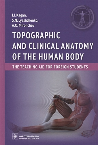
Kagan I., Lyashchenko S., Mironchev A. Topographic and clinical anatomy of the human body: the teaching aid for foreign students
Th is teaching aid is the additional source of information on topographical anatomy of regions of the human body and clinical anatomy of inner organs to the main textbook. Its English version is intended for students who take a training course in foreign faculty of the university. The teaching aid is illustrated by pictures, which are located on opening pages. On each such opening page, to the right on the odd page, there is the description of region or organ, and to the left on the even page, there is its location according to the anatomic region or organ picture. Such disposition makes the presentation of educational material more visual and makes the learning process easier.
2240 Руб.
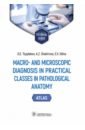
Цыплаков Дмитрий Эдуардович, Шакирова Ася Закиевна, Силина Екатерина Владимировна Macro- and microscopic diagnosis in practical classes in pathological anatomy
The tutorial guide offers a description of macro- and microreparations on general and special pathological anatomy studied in practical classes, provided with corresponding illustrations. The authors provide an overview of the principles of making macropreparations and the basics of histological technique and demonstrate staining histological sections and methods of histochemical, ultrastructural and immunohistochemical studies. The guide provides brief anatomical and histological characteristics of normal organs and a pattern of macropreparation description as well as the procedure of studying micropreparations. The tutorial guide contains 348 drawings, including photographs of macroand microreparations. The tutorial guide is composed in accordance with the curriculum for pathological anatomy course and intended for students receiving instruction in English for the specialty 31.05.01 "General Medicine".
3730 Руб.
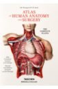
Bourgery J. M., Jacob N. M. Atlas of Human Anatomy and Surgery. The Complete
Discover one of the most comprehensive and beautifully illustrated anatomical treatises ever published. The product of more than two decades of dedication, and spanning descriptive, surgical, microscopic, and general anatomy, as well as embryology, The Atlas of Anatomy marks to this day a major achievement in medical history, and a breathtaking insight into the workings and wonder of the human body.
5049 Руб.

8Pcs Skeleton Model Wholesale Learn Aid Anatomy Art Sketch Halloween Flexible Human Anatomical Anatomy Bone
296.6 Руб.
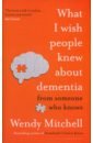
Mitchell Wendy, Wharton Anna What I Wish People Knew About Dementia. From Someone Who Knows
What can a diseased brain tell us about being human, living our own lives better and helping those with dementia get the best from theirs? When Wendy Mitchell was diagnosed with young-onset dementia at the age of fifty-eight, her brain was overwhelmed with images of the last stages of the disease - those familiar tropes, shortcuts and cliches that we are fed by the media, or even our own health professionals. But her diagnosis far from represented the end of her life. Instead, it was the start of a very different one. Wise, practical and life affirming, What I Wish People Knew About Dementia combines anecdotes, research and Wendy Mitchell's own brilliant wit and wisdom to tell readers exactly what she wishes they knew about dementia.
3500 Руб.
Mitchell Wendy What I Wish People Knew About Dementia
What can a diseased brain tell us about being human, living our own lives better and helping those with dementia get the best from theirs? When Wendy Mitchell was diagnosed with young-onset dementia at the age of fifty-eight, her brain was overwhelmed with images of the last stages of the disease - those familiar tropes, shortcuts and cliches that we are fed by the media, or even our own health professionals. But her diagnosis far from represented the end of her life. Instead, it was the start of a very different one. Wise, practical and life affirming, What I Wish People Knew About Dementia combines anecdotes, research and Wendy Mitchell's own brilliant wit and wisdom to tell readers exactly what she wishes they knew about dementia.
2519 Руб.
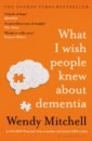
Anatomy for the Artist
Unlock your inner artist and learn how to draw the human body in this beautifully illustrated art book by celebrated artist and teacher Sarah Simblet. This visually striking guide takes a fresh approach to drawing the human body. A combination of innovative photography and drawings, practical life-drawing lessons, and in-depth explorations of the body’s surface and underlying structure are used to reveal and celebrate the human form. Combining specially-commissioned photographs of models with historical and contemporary works of art and her own dynamic life drawing, Sarah leads us inside the human body to map its skeleton, muscle groups, and body systems. Detailed line drawings superimposed over photographs reveal the links between the body’s appearance and its construction. Six drawing classes show how to observe different parts of the body and give expert guidance on how to draw them. Inspirational master classes on famous works, ranging from a Michelangelo study to a Degas painting, show how artists have depicted the human body over the centuries. Each master class includes a photograph of a model holding the same pose as in the painting, to highlight details of anatomy and show how the artist has interpreted them. Understanding anatomy is the key to drawing the human body successfully. As well as being the perfect reference, Anatomy for the Artist will inspire you to find a model, reach for your pencil, and start drawing.
1800 Руб.
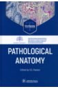
Pathological Anatomy. Textbook
This textbook is a contribution by a community of experienced lecturers from the Academician A.I. Strukov Department of Pathological Anatomy, I.M. Sechenov First Moscow State Medical University (Sechenov University), among contributors there are also leading experts from other medical universities. Coverage of the main issues of general and special pathological ana tomy is based on the principle of unity between structure and function. The authors tried to show not only the morphological basis of pathological processes and diseases, but also their connection with changes in body functions, resulting in certain symptoms of diseases, as well as the dynamics of their development. This approach provides a basis for further study of all clinical disciplines. Besides pathological anatomy, the manual describes general medical concepts, rules of writing clinical and pathoanatomic diagnoses, categories and causes of diagnostic discrepancies, etc. It helps students develop clinical thinking possible only if based on deep knowledge and understanding of objective laws that underlie pathological processes in the affected body. Thus, the study of pathological anatomy is the most important stage in training clinicians and this text meets the challenge. The textbook is intended for students of medical, pediatric, preventive medicine and dental facul ties of medical universities.
6898 Руб.
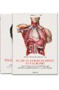
Le Minor Jean-Marie, Sick Henri Bourgery. Atlas of Human Anatomy and Surgery
We owe a great debt to Jean Baptiste Marc Bourgery (1797–1849) for his Atlas of Anatomy, which was not only a massive event in medical history, but also remains one of the most comprehensive and beautifully illustrated anatomical treatises ever published in any language. In 1830, having received his doctorate in medicine three years prior, Bourgery began work on his magnificent atlas in cooperation with illustrator Nicolas Henri Jacob (1782–1871), a student of the French painter Jacques Louis David. The first volumes were published the following year, but completion of the treatise required nearly two decades of dedication; Bourgery lived just long enough to finish his labor of love, but the last of the treatise’s eight volumes was not published in its entirety until five years after his death. The four parts of Bourgery’s treatise cover descriptive anatomy, surgical anatomy and techniques (exploring in detail nearly all the major operations that were performed during the first half of the 19th century), general anatomy and embryology, and microscopic anatomy. Jacob’s spectacular hand-colored, life-size lithographs are remarkable for their clarity, color, and aesthetic appeal, reflecting a combination of direct laboratory observation and illustrative research; the images are to this day unsurpassed in anatomical illustration.
3864 Руб.
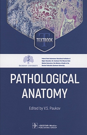
Paukov V.S. Pathological Anatomy: textbook
This textbook is a contribution by a community of experienced lecturers from the Academician A.I. Strukov Department of Pathological Anatomy, I.M. Sechenov First Moscow State Medical University (Sechenov University), among contributors there are also leading experts from other medical universities. Coverage of the main issues of general and special pathological ana tomy is based on the principle of unity between structure and function. The authors tried to show not only the morphological basis of pathological processes and diseases, but also their connection with changes in body functions, resulting in certain symptoms of diseases, as well as the dynamics of their development. This approach provides a basis for further study of all clinical disciplines. Besides pathological anatomy, the manual describes general medical concepts, rules of writing clinical and pathoanatomic diagnoses, categories and causes of diagnostic discrepancies, etc. It helps students develop clinical thinking possible only if based on deep knowledge and understanding of objective laws that underlie pathological processes in the affected body. Thus, the study of pathological anatomy is the most important stage in training clinicians and this text meets the challenge. The textbook is intended for students of medical, pediatric, preventive medicine and dental facul ties of medical universities.
5507 Руб.