bronchial tree model lungs medical teaching tool anatomical transparent science human anatomy
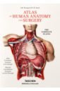
Bourgery J. M., Jacob N. M. Atlas of Human Anatomy and Surgery. The Complete
Discover one of the most comprehensive and beautifully illustrated anatomical treatises ever published. The product of more than two decades of dedication, and spanning descriptive, surgical, microscopic, and general anatomy, as well as embryology, The Atlas of Anatomy marks to this day a major achievement in medical history, and a breathtaking insight into the workings and wonder of the human body.
5049 Руб.
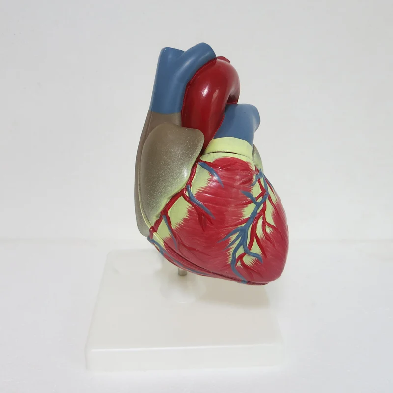
3 times big PVC Cardiac anatomy model Medical teaching tool instructional tool Clinic Figurines
1761.04 Руб.
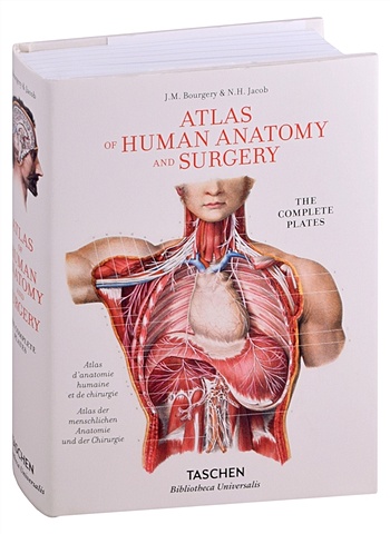
Bourgery J.M., Jacob N.H. Atlas of Human Anatomy and Surgery
We owe a great debt to Jean Baptiste Marc Bourgery (1797-1849) for his Atlas of Anatomy, which was not only a massive event in medical history, but also remains one of the most comprehensive and beautifully illustrated anatomical treatises ever published.Bourgery began work on his magnificent atlas in 1830 in cooperation with illustrator Nicolas Henri Jacob (1782-1871), a student of the French painter Jacques Louis David. The first volumes were published the following year, but completion of the treatise required nearly two decades of dedication; Bourgery lived just long enough to finish his labor of love, but the last of the treatise's eight volumes was not published in its entirety until five years after his death.The eight volumes of Bourgery's treatise cover descriptive anatomy, surgical anatomy and techniques (exploring in detail nearly all the major operations that were performed during the first half of the 19th century), general anatomy and embryology, and microscopic anatomy. Jacob's spectacular hand-colored lithographs are remarkable for their clarity, color, and aesthetic appeal, reflecting a combination of direct laboratory observation and illustrative research. Unsurpassed to this day, the images offer exceptional anatomical insight, not only for those in the medical field but also for artists, students, and anyone interested in the workings and wonder of the human body.
2471 Руб.
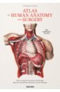
Bourgery J. M., Jacob N. H. Atlas of Human Anatomy and Surgery
We owe a great debt to Jean Baptiste Marc Bourgery (1797-1849) for his Atlas of Anatomy, which was not only a massive event in medical history, but also remains one of the most comprehensive and beautifully illustrated anatomical treatises ever published. Bourgery began work on his magnificent atlas in 1830 in cooperation with illustrator Nicolas Henri Jacob (1782-1871), a student of the French painter Jacques Louis David. The first volumes were published the following year, but completion of the treatise required nearly two decades of dedication; Bourgery lived just long enough to finish his labor of love, but the last of the treatise's eight volumes was not published in its entirety until five years after his death. The eight volumes of Bourgery's treatise cover descriptive anatomy, surgical anatomy and techniques (exploring in detail nearly all the major operations that were performed during the first half of the 19th century), general anatomy and embryology, and microscopic anatomy. Jacob's spectacular hand-colored lithographs are remarkable for their clarity, color, and aesthetic appeal, reflecting a combination of direct laboratory observation and illustrative research. Unsurpassed to this day, the images offer exceptional anatomical insight, not only for those in the medical field but also for artists, students, and anyone interested in the workings and wonder of the human body.
15147 Руб.
Bourgery J.M., Jacob N.H. Atlas of Human Anatomy and Surgery
We owe a great debt to Jean Baptiste Marc Bourgery (1797-1849) for his Atlas of Anatomy, which was not only a massive event in medical history, but also remains one of the most comprehensive and beautifully illustrated anatomical treatises ever published.Bourgery began work on his magnificent atlas in 1830 in cooperation with illustrator Nicolas Henri Jacob (1782-1871), a student of the French painter Jacques Louis David. The first volumes were published the following year, but completion of the treatise required nearly two decades of dedication; Bourgery lived just long enough to finish his labor of love, but the last of the treatise's eight volumes was not published in its entirety until five years after his death.The eight volumes of Bourgery's treatise cover descriptive anatomy, surgical anatomy and techniques (exploring in detail nearly all the major operations that were performed during the first half of the 19th century), general anatomy and embryology, and microscopic anatomy. Jacob's spectacular hand-colored lithographs are remarkable for their clarity, color, and aesthetic appeal, reflecting a combination of direct laboratory observation and illustrative research. Unsurpassed to this day, the images offer exceptional anatomical insight, not only for those in the medical field but also for artists, students, and anyone interested in the workings and wonder of the human body.
2471 Руб.
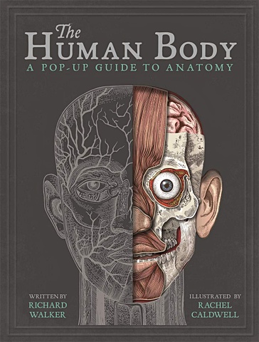
Уокер Р. The Human Body
It's 1839 and you are a medical student working on your first human body dissection! Under the watchful eye of Dr Walker, peel the flaps back to reveal the inner workings of the human body, from bone and muscle, to the brain, eyes, heart, lungs and everything in-between. Victorian-inspired illustrations meet with medical notes and sketches to give a complete in-depth exploration of how the human body works.
4488 Руб.
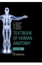
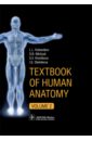
Колесников Лев Львович, Никитюк Дмитрий Борисович, Клочкова С. В. Textbook of Human Anatomy. In 3 vol. Volume 1. Locomotor apparаtus
This book provides essential facts of human anatomy for medical students. It demonstrates the basic knowledge for exam preparation and practice review of visual experiences. Plenty of clear illustrations (more than 900 pictures, radiographic and cross-sectional images) help students memorize the topics of anatomy. Modern imaging technologies allow the depiction of organs and systems in a variety of ways to gain thorough knowledge and to link to the clinical setting. The content of the book corresponds to the Federal Program for medical education. Material is divided according to systemic anatomy into three volumes. Volume 1 contains Introduction, Human development, and Locomotor apparatus.
4999 Руб.
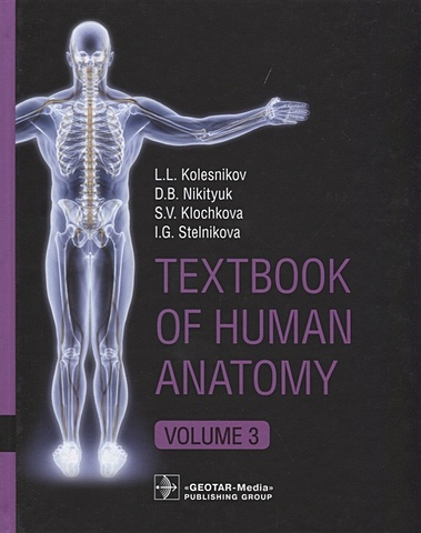
Колесников Л., Никитюк Д., Клочкова С., Стельникова И. Textbook of Human Anatomy. In 3 volume. Volume 3. Nervous system. Esthesiology/Анатомия человека. Учебник на английском языке в трех томах. Том 3
This book provides essential facts of human anatomy for medical students. It demonstrates the basic knowledge for exam preparation and practice review of visual experiences. Plenty of clear illustrations (more than 900 pictures, radiographic and cross-sectional images) help students memorize the topics of anatomy.
3249 Руб.

8Pcs Skeleton Model Wholesale Learn Aid Anatomy Art Sketch Halloween Flexible Human Anatomical Anatomy Bone
296.6 Руб.
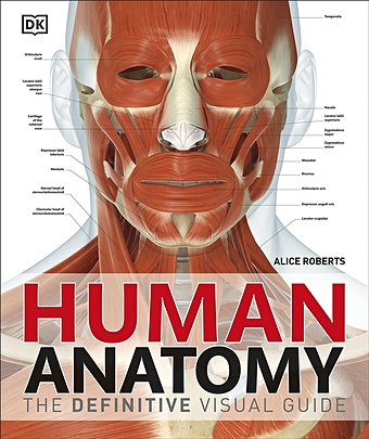
Human Anatomy
The definitive visual guide to human anatomy Discover fascinating facts about the human body in Human Anatomy. Did you know that your stomach is only a centimetre away from the bottom of your heart? You'll get to see all the biological wonders of the human body, often at life-size, with spectacular anatomical images showing the body's structure in incredible detail, from bone sutures to lymph nodes. Professor Alice Roberts takes you on a journey through all the body's systems, working down from the head to the toes, with a clear overview of each system. Amazing body diagrams and exhaustive annotations give you all the key details on organs and body structures. Human Anatomy is perfect for students of human biology and physiology with the quality of digital images and level of detail making it an invaluable study resource. Or if you are just curious to learn how your body works, this anatomy atlas is the perfect guide for you.
2572 Руб.
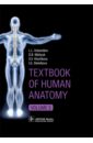
Колесников Лев Львович, Никитюк Дмитрий Борисович, Клочкова Светлана Валерьевна, Стельникова Ирина Textbook of Human Anatomy. In 3 volumes. Volume 2. Splanchnology and cardiovascular system
This book provides essential facts of human anatomy for medical students. It demonstrates the basic knowledge for exam preparation and practice review of visual experiences. Plenty of clear illustrations (more than 900 pictures, radiographic and cross-sectional images) help students memorize the topics of anatomy. Modern imaging technologies allow the depiction of organs and systems in a variety of ways to gain thorough knowledge and to link to the clinical setting. The content of the book corresponds to the Federal Program for medical education. Material is divided according to systemic anatomy into three volumes. Volume 2 contains information about internal organs development (Respiratory, Urinary, Reproductive, Lymphoid, Cardiovascular systems and Endocrine glands).
4538 Руб.
Колесников Лев Львович, Никитюк Дмитрий Борисович, Клочкова Светлана Валерьевна Textbook of Human Anatomy. Volume 3. Nervous system
This book provides essential facts of human anatomy for medical students. It demonstrates the basic knowledge for exam preparation and practice review of visual experiences. Plenty of clear illustrations (more than 900 pictures, radiographic and cross-sectional images) help students memorize the topics of anatomy. Modern imaging technologies allow the depiction of organs and systems in a variety of ways to gain thorough knowledge and to link to the clinical setting. The content of the book corresponds to the Federal Program for medical education. Material is divided according to systemic anatomy into three volumes. Volume 3 contains information about nervous system structure (Central, Peripheral, Autonomic nervous systems) and sense organs.
3630 Руб.
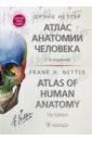
Неттер Фрэнк Атлас анатомии человека. Терминология на русском, латинском и английском языках
"Атлас анатомии" Фрэнка Неттера вот уже более 30 лет является лучшим и самым популярным пособием по анатомии человека для студентов-медиков во всем мире. Акцент сделан на клиническом значении анатомии, что дает возможность изучить строение тела человека в таком ракурсе, который нужен для последующего становления в профессии врача. Атлас представляет собой перевод седьмой, значительно обновленной и дополненной версии классического издания д-ра Неттера. Включено более 100 новых иллюстраций, пополнен ряд рентгенологических, КТ- и МРТ-изображений, полученных благодаря новым, усовершенствованным технологиям. В дополнение к справочным таблицам по мышцам добавлены таблицы с областями наибольшего клинического значения из-за распространенности повреждений в этих частях тела. В новом издании термины приведены на трех языках - русском, латинском и английском, все основные материалы книги, включая таблицы, дублируются на русском и английском языках. Такой подход поможет читателю овладеть знаниями анатомической терминологии в полном объеме, а также даст возможность подготовить себя к чтению литературы на иностранных языках. Вся терминология соответствует Международной анатомической терминологии (Terminologia Anatomica). Издание предназначено студентам медицинских вузов, в том числе обучающимся на иностранных языках, в качестве незаменимого пособия при изучении как нормальной, так и топографической анатомии. Frank Netter's "Atlas of Human Anatomy" has been the best and most popular human anatomy manual for medical students worldwide for more than 30 years. It emphasizes the clinical significance of anatomy, making it possible to study the structure of the human body in perspective necessary for subsequent development in the medical profession. The atlas provides a translation of the seventh, significantly updated and expanded version of Dr. Netter's classic edition. It includes more than 100 new illustrations, the set of X-ray, CT and MRI images is enriched thanks to new, improved technologies. Besides reference tables for muscles, there are tables of most clinically significant areas covering the most common injuries these parts of the body are subjected to. In the new edition, the terms are given in three languages - Russian, Latin and English. All main materials of the book, including tables, are duplicated in Russian and English. This approach will help the reader to master the knowledge of anatomical terminology most fully and gives a chance to get ready for reading in foreign languages. All terminology meets the requirements of the International Anatomical Terminology (Terminologia Anatomica). The edition is an invaluable resource for medical students, including those studying in foreign languages, in their study of both normal and topographic anatomy. 7-е издание.
18852 Руб.